IVIS Spectrum - System specifications
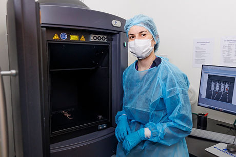
IVIS Spectrum
CCD camera
Back-thinned, back-illuminated Grade 1 CCD sensor
Thermoelectric cooling to -90°C for low dark current and noise
Pixel size: 13.5 µm
Minimum pixel resolution: 20 µm
Sensor dimensions: 2048 × 2048 pixels
Maximum sensor binning: 16
Quantum efficiency: 85% in the 500–700 nm range and 30% in the 400–900 nm range
Lens
6-inch diameter optics
Numerical aperture adjustable from f/1 to f/8
Discrete magnifications: 1.5×, 2.5×, 5×, and 8.7×
Corresponding fields of view: 3.9 cm to 22.5 cm
High-resolution imaging down to 20 µm with small field of view
Optical path
High-throughput imaging with a large field of view (up to 5 mice or 2 rats simultaneously)
10 high-efficiency narrow bandpass excitation filters (415–760 nm, 30 nm bandwidth)
18 high-efficiency narrow bandpass emission filters (490–850 nm, 20 nm bandwidth)
Optical switch enabling both epi-illumination and trans-illumination fluorescence modes
Imaging chamber
Temperature-controlled platform for maintaining physiological temperature during imaging
Integrated RAS-4 gas anesthesia system
light tight chamber
Software
Radiometric calibration of measurements in radiance (bioluminescence) and radiance efficiency (fluorescence) for reproducible results
Spectral unmixing for separating multiple fluorescent or bioluminescent signals and background subtraction
Co-registration with anatomical atlases or CT/MRI for overlaying structural and functional data
3D bioluminescence and fluorescence tomography with depth estimation and quantitative analysis
Exportation of DICOM image format for 3D co-registration
Available accessories
Fluorescence and bioluminescence phantom mouse for calibration, validation, quality control
Mouse Imaging Shuttle (MIS) for 3D multimodal co-registration with microCT or MRI systems
Operational protocols and risk assessments
The SOPs and RAs related to the use of the IVIS Spectrum system are available https://drive.google.com/drive/u/1/folders/1yU5AkpK4jt7LfB_xHxhKC4837ZLwxuR2, upon request. These documents are stored in the designated folder and include:
IVIS Spectrum user manual
SOP for IVIS Spectrum utilization
SOP for RAS-4 rodent anesthesia system
RA for IVIS Spectrum with anesthesia
Revvity useful information