SkyScan 1276 - Main applications
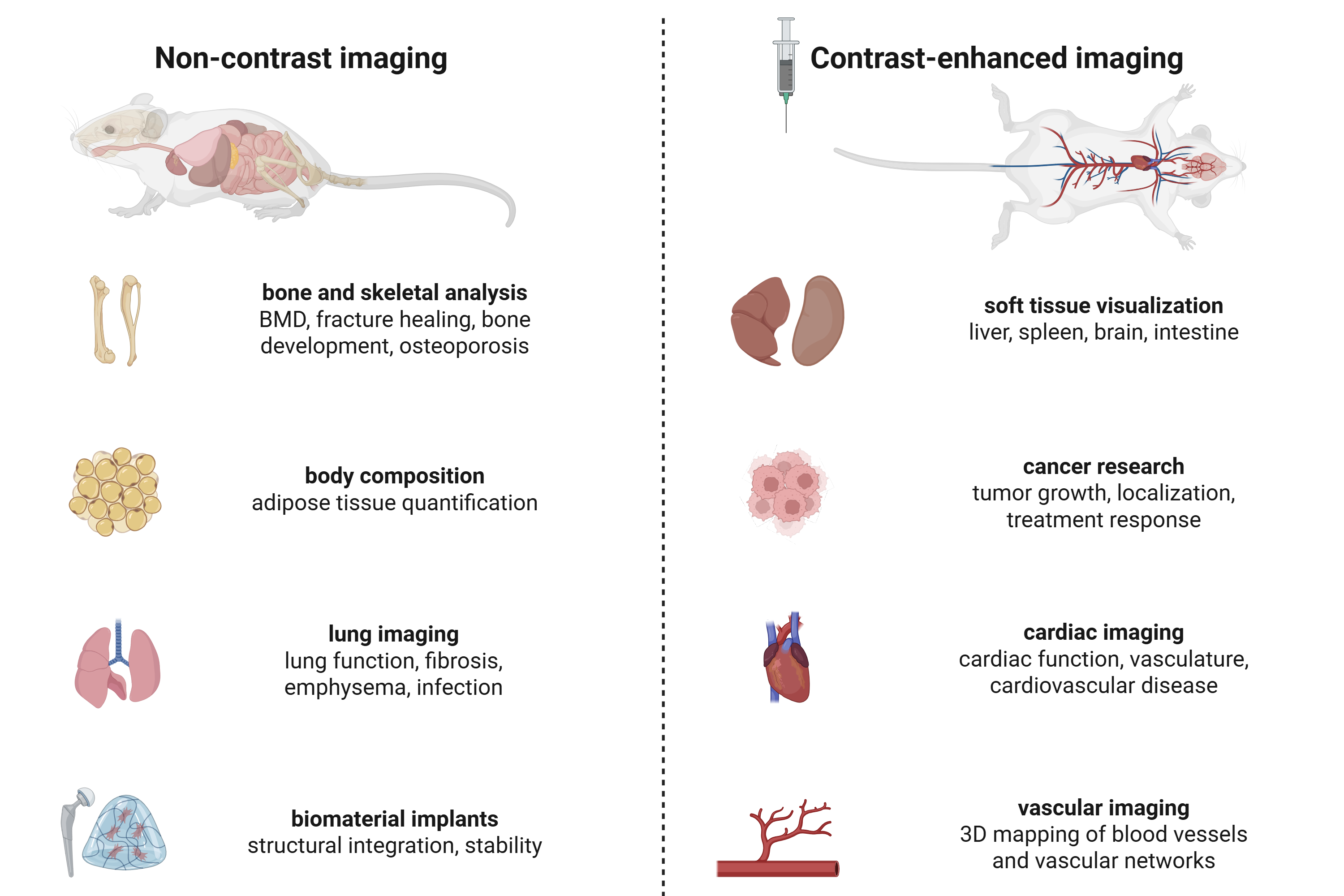
The main application of the SkyScan 1276 is non-invasive in vivo micro-computed tomography (microCT) imaging for visualizing anatomical structures in small animal models with high spatial resolution. Its ability to capture detailed 3D images of bone and soft tissue, combined with fast scanning speeds and low radiation doses, makes it especially powerful for longitudinal studies of disease progression, treatment response, and physiological changes over time in live animals.
In vivo microCT imaging
Common applications of in vivo microCT
Here are common applications of in vivo microCT imaging:
- Bone morphometry
Analysis of bone microarchitecture and bone mineral density (BMD)
Studies of osteoporosis, bone healing, growth, and regeneration
- Lung imaging
Functional respiratory analysis using respiratory gating (4D microCT)
Monitoring pulmonary disease progression (fibrosis, emphysema, tumor, tuberculosis)
- Body composition analysis
Quantification of adipose tissue
Quantification of muscular tissue
- Biomaterial implant evaluation
Assessment of biocompatibility, structural integrity, and stability of implants
- Soft tissue visualization (contrast agent)
Morphological and structural analysis of liver, spleen, and kidneys
Volumetric analysis of soft tissue organs
- Cancer research (contrast agent)
Monitoring tumor growth, localization, and vascularization
Evaluating efficacy of anti-cancer therapies
- Cardiac imaging (contrast agent)
Functional cardiac analysis with cardiac gating (4D microCT)
Tracking cardiovascular disease progression (atherosclerosis, aneurysms, stroke)
- Vascular imaging (contrast agent)
3D mapping and quantification of blood vessels and vascular networks
Additional microCT applications
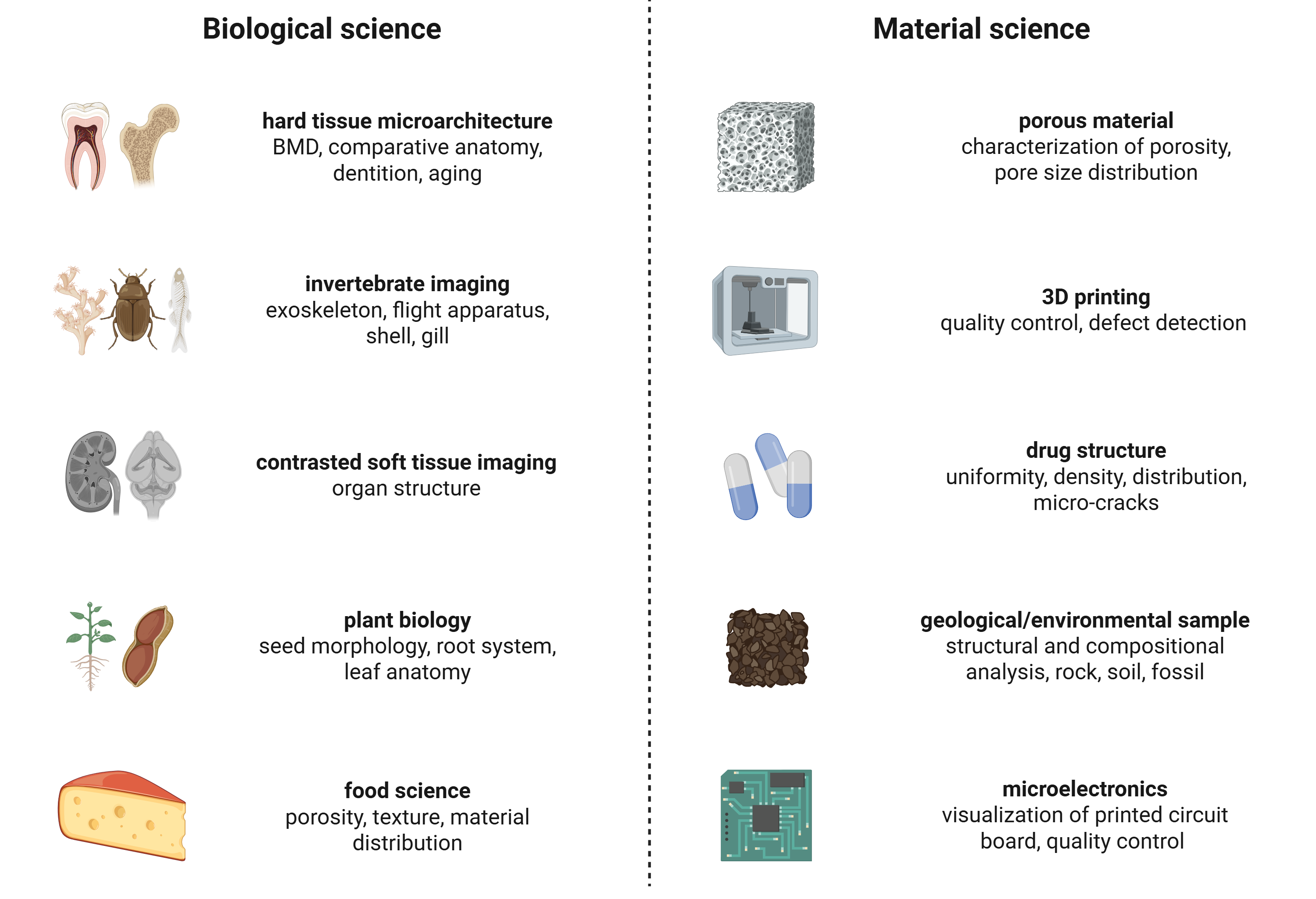
Common applications of microCT for biological and material science
Here are some biological research applications of microCT imaging:
- Hard tissue micro-architecture
Comparative anatomy studies
Dental structure and development
Aging-related skeletal changes
- Invertebrate imaging
Visualization of exoskeletons, flight apparatus, shells, and gills
- Contrast-enhanced soft tissue imaging
High-resolution visualization of internal organ structures
- Plant biology
Seed morphology and development
Root system architecture
Leaf anatomy and venation patterns