Ultima 2Pplus - Main applications
The Ultima 2Pplus two-photon microscope is designed to enable high-resolution, intravital, cellular and subcellular imaging in anesthetized or head-fixed awake animals, including during controlled sensory stimulation or task performance. It provides optical sectioning and deep-tissue imaging with reduced phototoxicity, allowing precise spatial mapping of neural, immune, and vascular activity within intact brain and peripheral tissues.
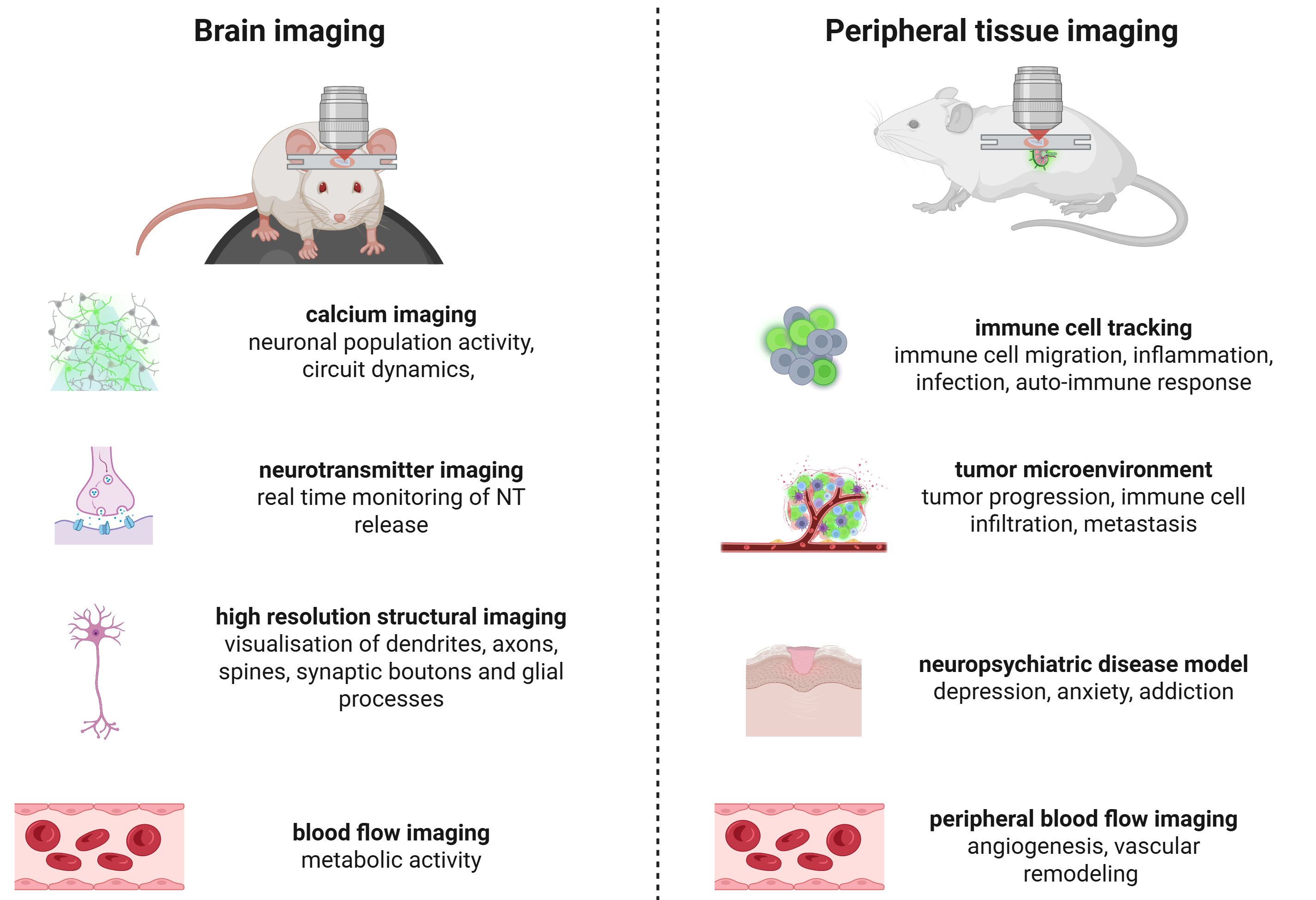
Common applications of intravital two-photon microscopy
Brain imaging
- Calcium imaging (GCamp, RCamp)
Enables optical detection of action potential–associated intracellular Ca²⁺ transients in single neurons and neuronal populations.
Supports imaging of excitatory and inhibitory circuits in cortical and subcortical regions during sensory stimulation, behavior, learning, rest, or spontaneous activity.
Allows quantification of firing dynamics, population synchrony, and circuit-level coding.
- Neurotransmitter imaging (dLight, GRAB, iGluSnFR)
Measures extracellular dynamics of neurotransmitters such as dopamine, glutamate, acetylcholine, and serotonin in vivo.
Enables direct assessment of neuromodulatory signaling and its relationship to neural activity.
Applied in studies of motivation, reinforcement learning, decision-making, attention, and neuropsychiatric disease models.
- Sequential multi-signal imaging
Allows imaging of multiple fluorescent reporters across sequential acquisitions or experimental sessions using a single excitation laser.
Enables comparison of activity across distinct neuronal populations, signaling pathways, or molecular readouts within the same brain region.
Suitable for static/static and static/dynamic experimental designs.
- Longitudinal circuit and ensemble tracking
Supports stable chronic imaging of the same neuronal populations over days to weeks through cranial windows.
Enables investigation of learning-induced plasticity, memory consolidation, recovery after injury, and neurodegenerative disease progression.
Facilitates analysis of neuronal identity retention and population-level stability or reorganization.
- Neurovascular and blood flow imaging
Enables visualization of cerebral vasculature, vessel diameter changes, and capillary blood flow using fluorescent tracers.
Supports studies of neurovascular coupling, metabolic demand, and vascular dysfunction.
Applied in aging, stroke, traumatic brain injury, and neurodegenerative disease research.
- Structural and synaptic imaging
High-resolution visualization of dendrites, axons, spines, synaptic boutons, and glial processes.
Allows correlation of functional activity with structural remodeling and synaptic plasticity.
Applied in developmental neuroscience, experience-dependent plasticity, and disease models.
Peripheral tissue imaging
Intravital imaging of peripheral organs and tissues requires direct optical access via implanted or surgically prepared imaging windows, enabling real-time visualization of cellular dynamics, signaling events, and microenvironmental interactions under physiological conditions.
- Immune cell tracking and interaction analysis
Visualizes immune cell migration, arrest, and cell–cell interactions in tissues such as skin, lymph nodes, lung, liver, and intestine.
Enables investigation of immune surveillance, inflammation, infection, and autoimmune responses.
Supports longitudinal tracking of immune behavior during disease progression or therapeutic intervention.
- Tumor and tumor microenvironment imaging
Enables high-resolution imaging of cancer cells, stromal components, and immune infiltrates in vivo.
Allows analysis of tumor cell invasion, metastasis, and cell–cell interactions within the tumor microenvironment.
Applied in preclinical oncology and therapeutic response studies.
- Metabolic and biosensor imaging
Supports imaging of genetically encoded or dye-based biosensors reporting on metabolism, redox state, ion concentration, or intracellular signaling pathways.
Enables investigation of tissue stress, metabolic heterogeneity, and signaling dynamics in health and disease.
- Tissue remodeling and regeneration
Enables longitudinal imaging of cell migration, extracellular matrix remodeling, and tissue repair processes.
Applied in regenerative medicine, fibrosis, chronic inflammation, and aging research.
- Microvascular and blood flow imaging
Visualizes capillary perfusion, leukocyte trafficking, angiogenesis, and vascular remodeling in peripheral organs.
Enables studies of ischemia, inflammation, wound healing, and vascular pathology.
Supports assessment of tissue perfusion and vascular responses to pharmacological or genetic manipulation.